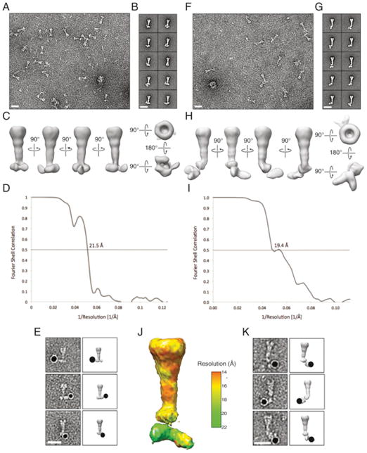
Figure 6. Two Conformations of the Tse6 Secretory Particle Revealed by Electron Microscopy.
(A and B) Addition of detergent dissociates EagT6 from the Tse6 secretion particle and causes a conformational change. (A) Coomassie-stained SDS-PAGE analysis and (B) representative class averages of purified Tse6-containing complex in the presence and absence of 0.03% β-D-dodecylmaltopyranoside.
(C) 3D density map and molecular fitting of the Tse6-Tsi6-VgrG1-EagT6-EF-TuPA complex. The identity of each subunit is indicated. The model for Tse6PAAR was generated using Phyre (Kelley and Sternberg, 2009).
(D) Tse6 requires EagT6 for intracellular accumulation. Western blot analysis of Tse6 levels in the indicated P. aeruginosa strains. RNA polymerase (RNAP) is used as a loading control.
(E) 3D density map and molecular fitting of the detergent-bound Tse6–Tsi6–VgrG1–EF-TuPA complex. Scale bars, 20 nm.
See also Figures S6 and S7 and Tables S2 and S3.

