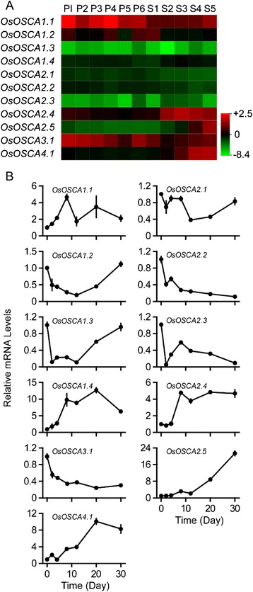
Fig. 4.

Expression profiles of OsOSCA genes during panicle and caryopsis development. a. The microarray data sets (GSE6893) of OsOSCA gene expression in organs at various developmental stages were reanalysed (Additional file 3: Table S3). The average log signal values of OsOSCA genes are presented in the form of a heat map. The colour key represents average log2 expression values of OsOSCA genes. The samples are indicated at the top of each lane. The following stages of panicle development are indicated as follows: P1, 0–3 cm; P2, 3–5 cm; P3, 5–10 cm; P4, 10–15 cm; P5, 15–22 cm; and P6, 22–30 cm. The following stages of caryopsis development are indicated as follows: S1, 0–2 dap (day after pollination); S2, 3–4 dap; S3, 5–10 dap; S4, 11–20 dap; and S5, 21–29 dap. A colour scale representing the average log signal values is shown on the right. b. The expression levels of OsOSCA genes during caryopsis development were monitored using qRT-PCR. Samples were collected at 0, 2, 4, 8, 12, 20, and 30 dap. The relative water content in corresponding stages is shown in Additional file 6: Figure S2. The relative mRNA levels of individual genes were normalised to that of actin. Error bars indicate the standard deviations (SD) of three biological replicates
