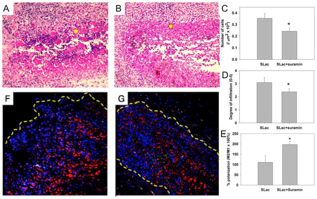
Figure 7.

In vivo evaluation of scaffolds. (A) Scaffolds with suramin-loading show large platelets of MDP uninfiltrated (*), compared to (B) a similar region (*) in SLac-only scaffolds. (C) Quantification of infiltration showed a significantly lower number of cells, (D) infiltrating loaded scaffolds to a diminished extent, (E) with a greater M2 macrophage polarization. Representative images of suramin-loaded (F) and -unloaded (G) scaffolds shown. Nuclei, DAPI blue; macrophages, CD68-red; M1, CCR7-green; M2, CD163-purple. (*, p < 0.01).
