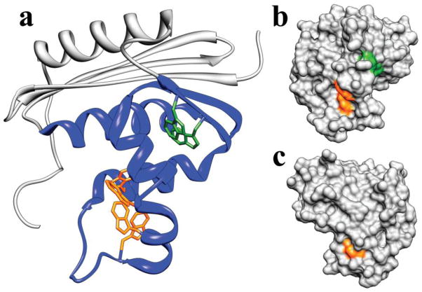
Figure 1.
Structure of E. coli RNase H. a. Ribbon diagram. Helices are labeled with letters and β-strands with Roman numerals. The region that is structured in the Icore intermediate is colored blue. Tryptophan residues are shown in stick (in the 4Trp variant, the two green tryptophans are mutated to phenylalanine, leaving only the four orange tryptophans). b. Surface contour of the RNase H crystal structure in a very similar orientation as in panel a. Only the tryptophan side chains are colored, to highlight the solvent exposure of these side chains in the native state. c. Structure in B rotated 180 degrees about the vertical axis. W104 on helix D is the only completely buried tryptophan.

