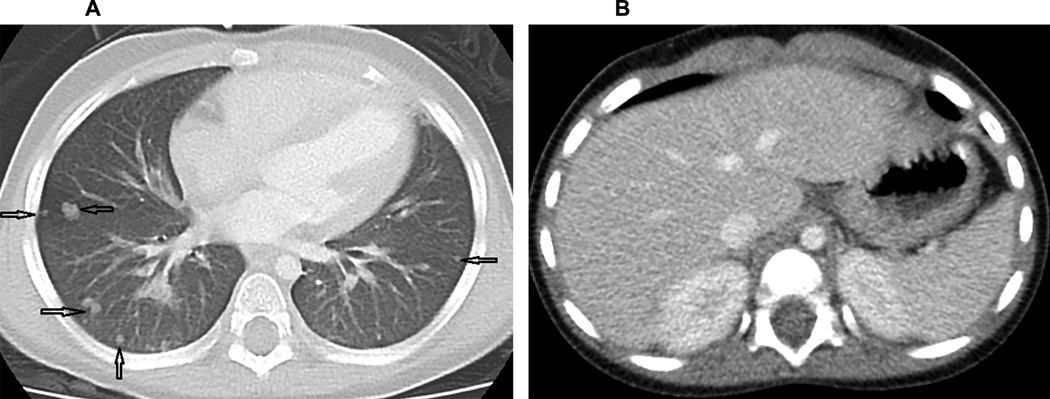
Figure 2.

Axial CT images through the lung bases (A) and abdomen (B). A. The chest image is displayed at a lung window/level, demonstrating multiple, bilateral, non-calcified pulmonary nodules of various sizes (arrows) B. A representative image through the upper abdomen shows uniform solid organs and absence of lesions in the liver, spleen and kidneys.
