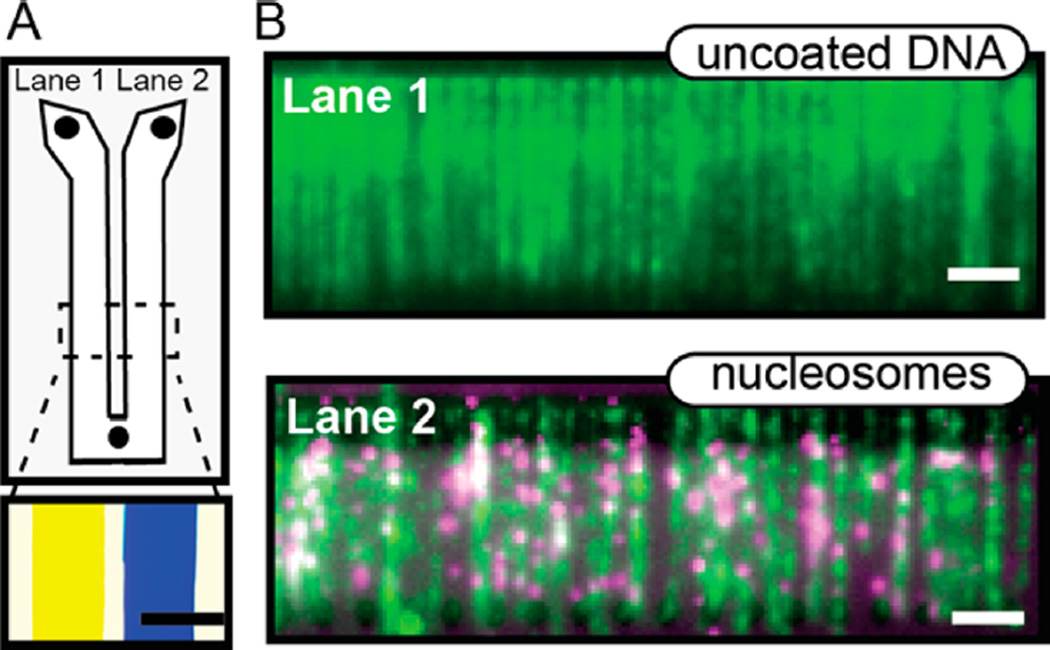
Figure 5.
A dual-lane flowcell for imaging two DNA substrates in the presence of buffer flow. (A) Cartoon schematic of the Y-shaped flowcell with two inlets and one outlet port. Each lane is 9 mm wide and separated by a 2 mm tape spacer (gray). Bottom panel: an image of yellow and blue food dye loaded into each of the two lanes. Scale bar: 5 mm. The lanes remain fluidically isolated for over 1 h. (B) Images captured from each lane during a single experiment. Lane 1 was assembled with λ-DNA, while lane 2 contained nucleosomecoated λ-DNA. Both channels were labeled with YOYO-1 DNA intercalating dye (Life Technologies). Nucleosomes were tagged with anti-FLAG QDs (magenta; 705 nm) and were exclusively observed in the right channel. Scale bar: 4 µm.

