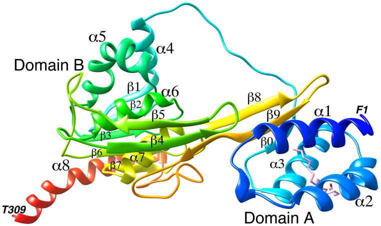
Figure 2. A ribbon diagram of the crystal structure of zebrafish IRBP module 1.
The N terminus is shown in blue and the C terminus in red. The domains and the secondary structure elements are marked. The oleic acid (OLA) binding site is shown with the bound oleic acid molecule. The terminal amino acids are also marked.

