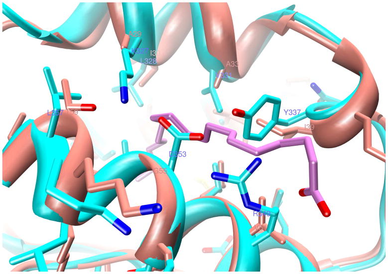
Figure 8. Homology modeling of module 2 (z2, in cyan) after z1 (in salmon color).
Several small-to-large side chain substitutions at the cavity perimeter as well as within the interior in domain A are depicted. The location of oleic acid C9=C10 cis double bond is marked by the labels 9 and 10. But for some structural rearrangements, these substitutions are therefore likely to eliminate this oleic acid binding site from z2.

