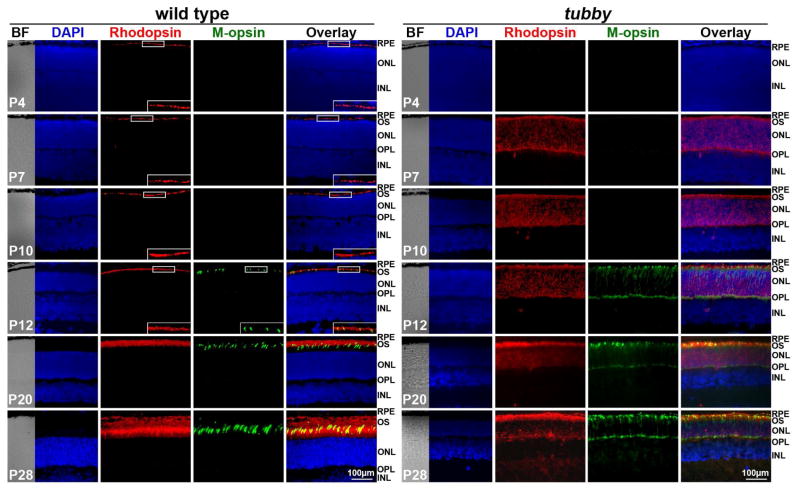
Figure 1.
Mislocalization of rhodopsin and M-opsin in tubby retinas. Immunocytochemistry of rhodopsin (red) and M-opsin (green) in wt (left) and tubby (right) retinas from P4 -P28 revealed that in wt retinas, rhodopsin is detectable at P4 and M-opsin at P12, whereas in tubby retinas, rhodopsin appears at P7 and M-opsin at P12 with mislocalization. Images shown are representative of 5–8 eyes per group. Nuclei were counterstained with DAPI. Inserts are the enlarged part of the boxed regions of the corresponding images. BF: bright field, RPE: retinal pigment epithelium, OS: outer segment, ONL: outer nuclear layer, OPL: outer plexiform layer, INL: inner nuclear layer. N=5–8, Scale bar, 100 μm.

