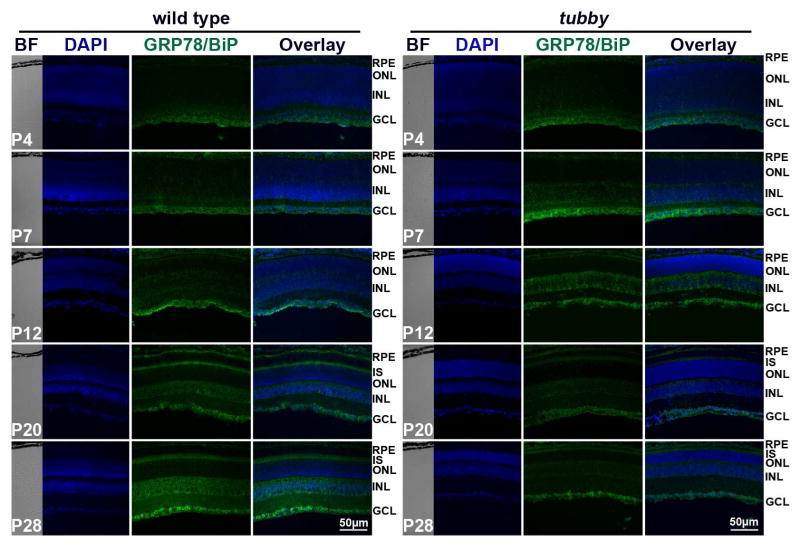
Figure 4.
Distribution and alteration of GRP78/BiP protein during retinal development. Immunocytochemistry showed that in wt and tubby retinas, GRP78/BiP is mainly expressed in the RPE, INL and GCL at P4 through P12 with expression being stronger in tubby retinas than the wt, but at P20 through P28, it was also expressed in the IS. However, tubby retinas have much less GRP78/BiP fluorescence compared to wt and to younger tubby retinas at P20 and P28. Representative images from each group are shown. BF: bright field, RPE: retinal pigment epithelium, IS: inner segment, ONL: outer nuclear layer, INL: inner nuclear layer, GCL: ganglion cell layer. N=8–10, Scale bar, 50 μm.

