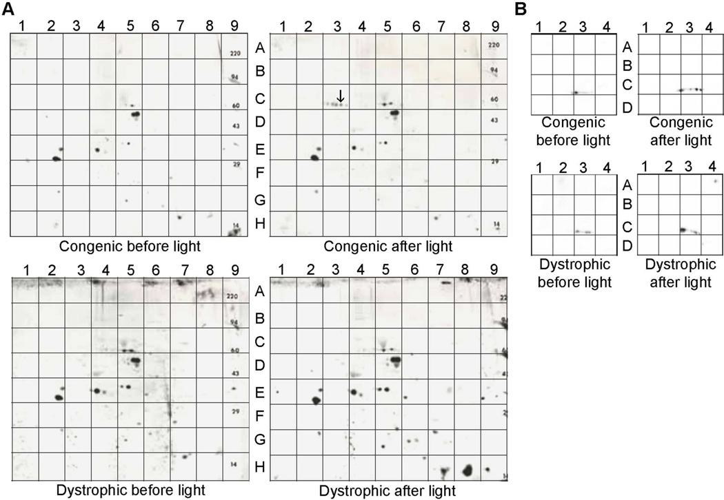
Fig. 1. Tyrosine-phosphorylated proteins present in RPE/choroid of 4 wk old RCS congenic and dystrophic rats.
(A) Protein homogenates were subjected to 2-D polyacrylamide-gel electrophoresis followed by western analysis to visualize anti-phosphotyrosine reactivity on blots from animals euthanized 1.0 h before light onset or 1.5 h after light onset. The spot denoted with an arrow in quadrant C3 of “congenic after light” corresponds to the location of the protein that was evaluated by MALDI-MS analysis (Table 1). (B) Regions A–D/1–4 of the blots in A) probed for anti-GDI1 immunoreactivity.

