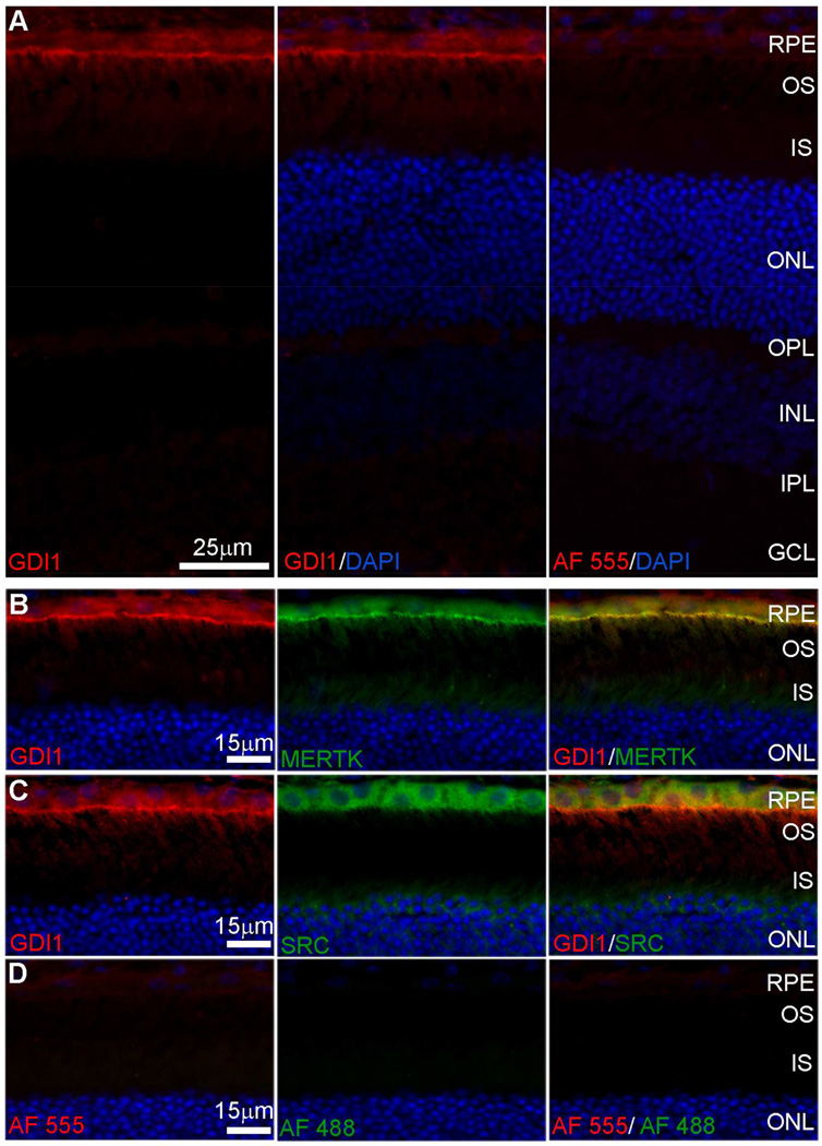
Fig. 2. Indirect fluorescence microscopy of GDI1 localization in cryosections of mouse retina/RPE/choroid.
(A) Anti-GDI1 immunoreactivity visualized using AlexaFluor 555 anti-mouse IgG (red) with DAPI staining of nuclei (blue). (B, C) High magnification images of the outer retina/RPE showing anti-GDI1 immunoreactivity (red) and (B) anti-Mertk immunoreactivity or (C) anti-SRC immunoreactivity visualized using AlexaFluor 488 anti-rabbit IgG (green). (D) Control sections from which primary antibodies were omitted. RPE, retinal pigment epithelium; OS, outer segments; IS, inner segments; ONL, outer nuclear layer; OPL, outer plexiform layer; INL, inner nuclear layer; IPL, inner plexiform layer; GCL, ganglion cell layer.

