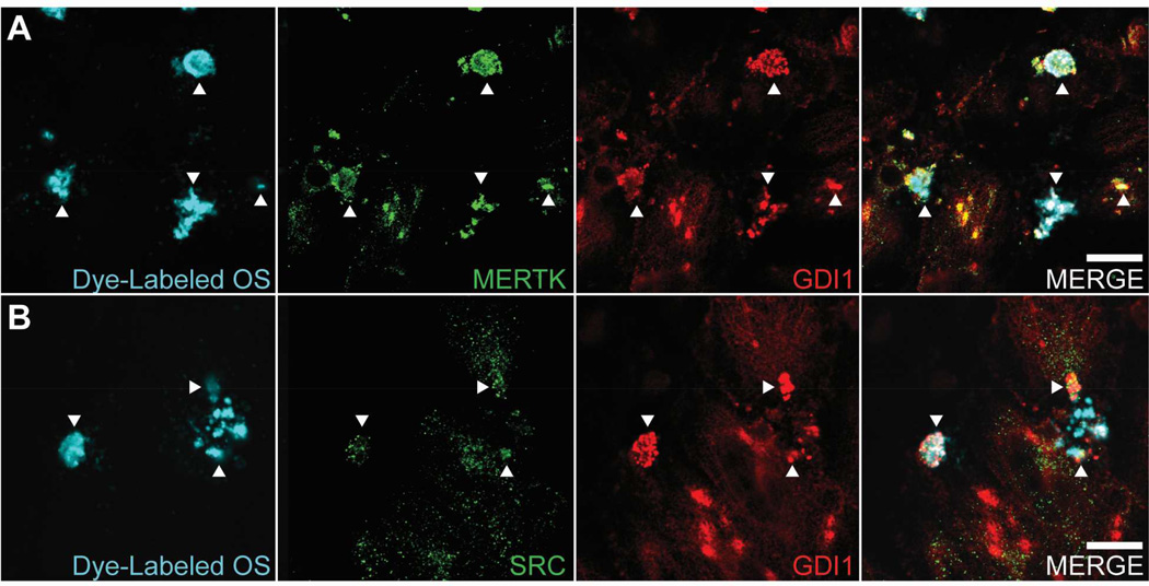
Fig. 7. Colocalization of GDI1 with MERTK and SRC on phagosomes.
Cultures of RPE-J cells were fed OS labeled with fluorescent dye (DyLight 405 nm) for 2 hours, fixed, immunostained for GDI1 and MERTK or GDI1 and SRC, and then subjected to indirect immunofluorescence confocal microscopy as in Fig. 6. (A) Panels from left to right show OS fluorescence, MERTK immunolabeling, GDI1 immunolabeling, and merge of OS, MERTK, and GDI1. (B) Panels from left to right show OS fluorescence, SRC immunolabeling, GDI1 immunolabeling, and merge of OS, SRC, and GDI1. Secondary antibodies were AlexaFluor-488 for the green channel and AlexaFluor-647 for the red channel. Controls which omitted dye-labeled OS, or MERTK/SRC antibody (green channel), or GDI1 antibody (red channel) confirmed the absence of spectral bleed-through between channels (see Supplemental Fig. 1). Arrowheads point to representative areas of colocalization for each antibody pair. All the colocalization seen is exclusively on the apical surface, with no colocalization apparent deeper in the cells, consistent with the association of GDI1 with MERTK and SRC on the plasma membrane. Scale bars are 10 µm.

