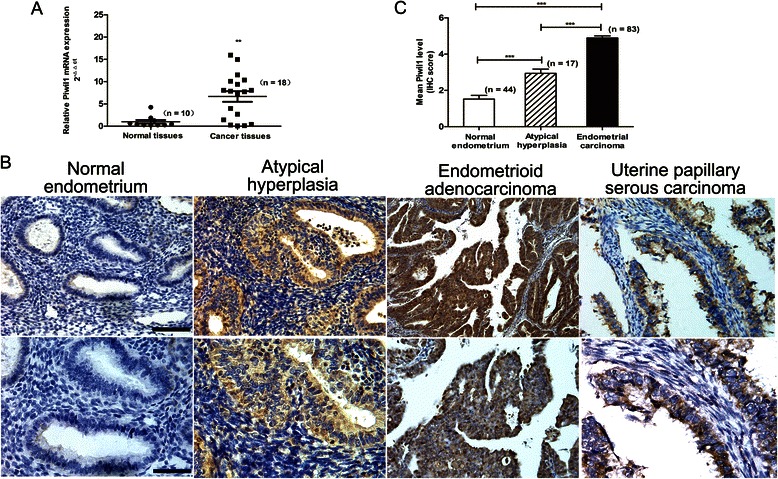
Fig. 1.

Expression of Piwil1 in human endometrial carcinoma tissue. a RT-qPCR analysis for Piwil1 mRNA in normal human endometrial tissues (n = 10) and endometrial cancer tissues (n = 18) (**P < 0.01). b Representative Piwil1 immunohistochemical staining of normal endometrial tissues, endometrial atypical hyperplasia and two endometrial cancer types. Original magnification 200×, scale bar, 100 μm (upper); 400×, scale bar, 50 μm (lower). c Immunohistochemistry scores (IS) of Piwil1 in normal endometrium (n = 44), atypical hyperplasia (n = 17), and endometrial carcinoma tissues (n = 83) (***P < 0.001)
