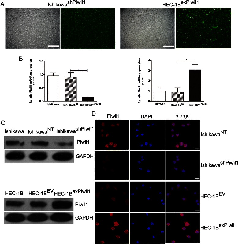
Fig. 2.

Overexpression or knockdown of Piwil1 in human endometrial cancer cell lines. a Stable transfection of Ishikawa cells with shRNA against Piwil1 and HEC-1B cells with Piwil1 expression plasmids. The percentage of transfected cells with fluorescence was > 95 %. (b and c) RT-qPCR and western blot demonstrated expression level of Piwil1 in Ishikawa, IshikawaNT and IshikawashPiwil1 cells or HEC-1B, HEC-1BEV and HEC-1BexPiwil1 cells (*P < 0.05). d Representative immunofluorescence images showing Piwil1 expression in IshikawaNT and IshikawashPiwil1 cells or in HEC-1BEV and HEC-1BexPiwil1 cells. Nuclei were stained with DAPI. Scale bars, 25 μm
