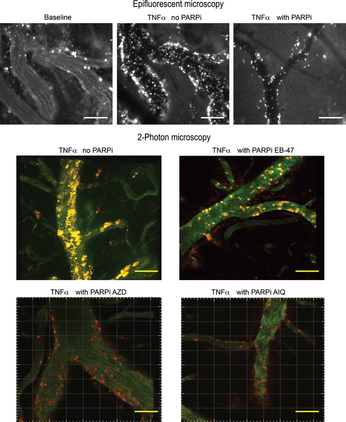
Fig. 4.

Images of pial vessels using intravital epifluorescent microscopy (upper panel) and 2-photon microscopy (lower panel). Animals were treated with TNFα (0.5 μg/mouse) with or without PARP-1 Inhibitor (PARPi) EB-47, AZD and AIQ (10 mg/kg). A reduction in the leukocyte adhesion to and migration across the blood brain barrier endothelium is observed. Vessels were stained with FITC-labeled Dextran 70 (green) and leukocytes were labeled with Rhodamine-6G (red/yellow color). Scale bars 100 μm
