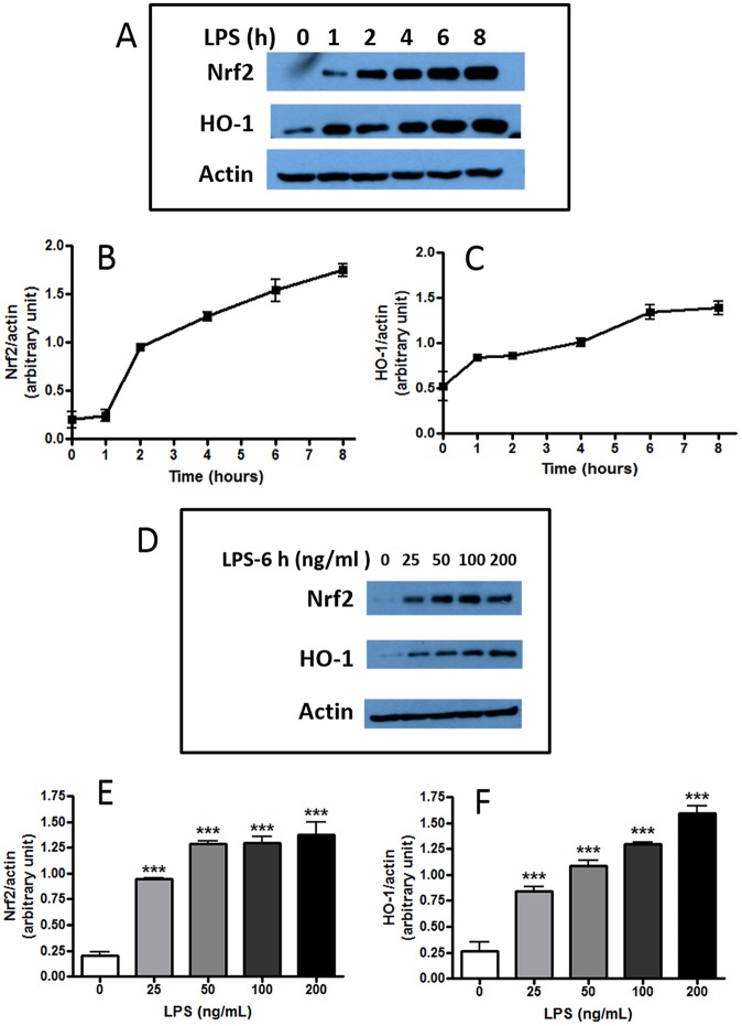
Fig 2. LPS induces Nrf2 and HO-1 protein expression in BV-2 microglial cells.
Confluent cells were serum starved for 4 h prior to stimulation with LPS (100 ng/ml) and assay for Nrf2 and HO-1 expression at different times. (A, B, C). Time- dependent increase in Nrf2 and HO-1 protein expression after stimulation with LPS (100 ng/ml). Results are means ± SEM of three independent experiments. (D, E, F) Dose-dependent increase of Nrf2 and HO-1 protein expression by LPS after a 6 h incubation time. Results are means ± SEM of three independent experiments and were analyzed by one-way ANOVA followed by Bonferroni post-tests. ***p<0.001 vs. no LPS control.

