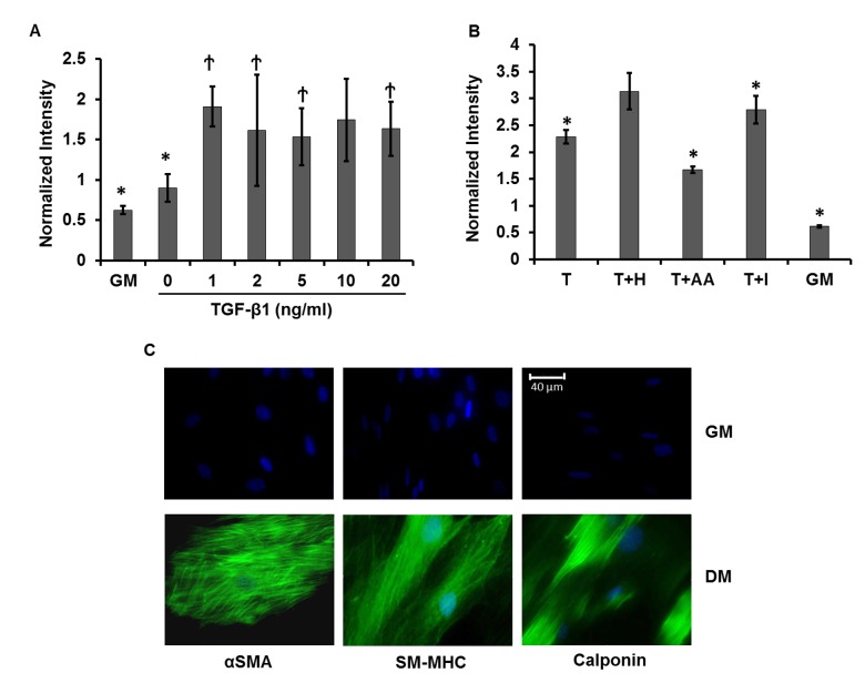
Fig 2. Optimal conditions for myogenic differentiation.
Optimized myogenic differentiation medium was determined by treating LVDP-pACTA2-ZsGreen transduced hBM-MSCs with (A) varying concentrations of TGF-β1 (0, 1, 2, 5, 10, and 20 ng/ml) or (B) a combination of 10 ng/ml TGF-β1 (T) and one of the following soluble factors: 30 μg/ml Heparin (H), 30 μM Ascorbic Acid (AA), or 2 μM Insulin (I). Red and green fluorescence intensities were measured by flow cytometry after 2 days and normalized intensity was shown. (A) * denotes p < 0.05 as compared to 10 ng/ml TGF-β1, Ϯ denotes p > 0.05 as compared to 10 ng/ml TGF-β1. (B) * denotes p < 0.05 as compared to T+H. (C) Immunostaining for SMC proteins (αSMA, smooth muscle myosin heavy chain (SM-MHC) and Calponin) under GM or the optimized differentiation medium (DM, T+H) for 7 days. Cell nuclei were counterstained with the Hoechst 33342. Images are representative of three independent experiments. Scale bar: 40 μm.

