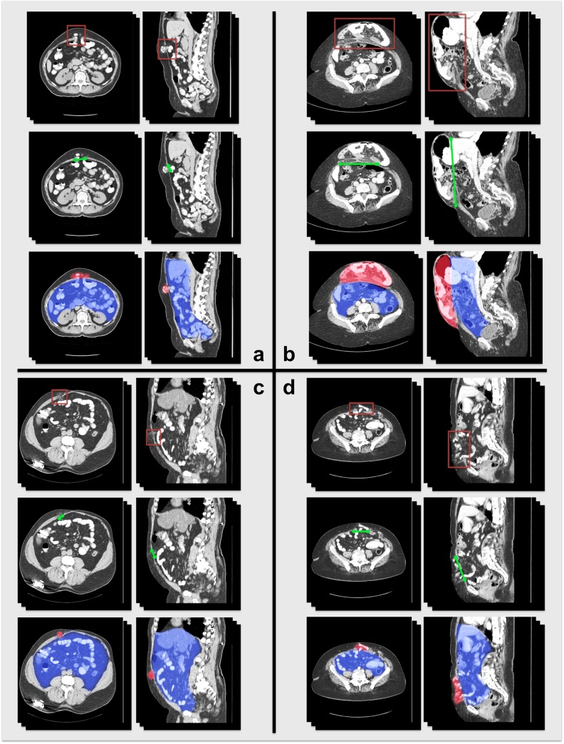
Fig 6. Illustration of VH characteristics on CT for four patients.
In each section, the first row illustrates the location of the VH; the second row illustrates the VH defect size at the anterior abdominal wall; the third row demonstrates the volume size of the hernia sac (red) and the abdominal cavity (blue).

