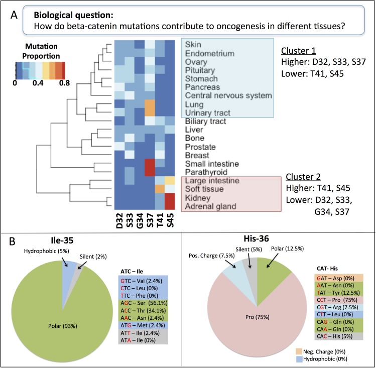
Fig 7. Mutation patterns of beta-catenin across cancer types.
(A) Hierarchical clustering of cancer tissues based on their pattern of mutations in the six most frequently mutated beta-catenin residues in cancer: Asp-32, Ser-33, Gly-34, Ser-37, Thr-41, and Ser-45. (Band C) Frequencies of the various possible amino acid substitutions at beta-catenin residues Ile-35 (B) and His-36 (C) observed in cancer samples. Amino acids are color coded and grouped in the pie charts according to the chemical nature of their side chains.

