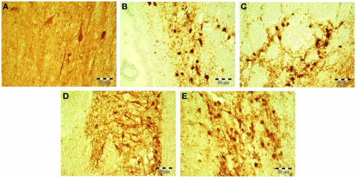
Figure 3.
(A–E) Calretinin-immunopostive neurons in the Ncl. medialis of a healthy control subject (A); Ncl. medialis of a patient with major depressive disorder (B); Ncl. lateralis of a patient with major depressive disorder (C); Ncl. medialis of a patient with schizophrenia (D); and Ncl. medialis of a healthy control subject (E). Scale bars correspond to 50 μm. Ncl., nucleus.

