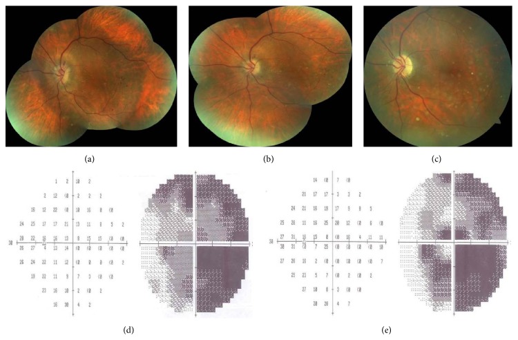
Figure 3.
Left eye, color fundus pictures taken at the first week (a), first month (b), and third month after the injection (c) showing the resolution of the disc edema with subsequent occurrence of pallor. Visual field test demonstrating the gradual improvement at the first (d) and third month (e).

