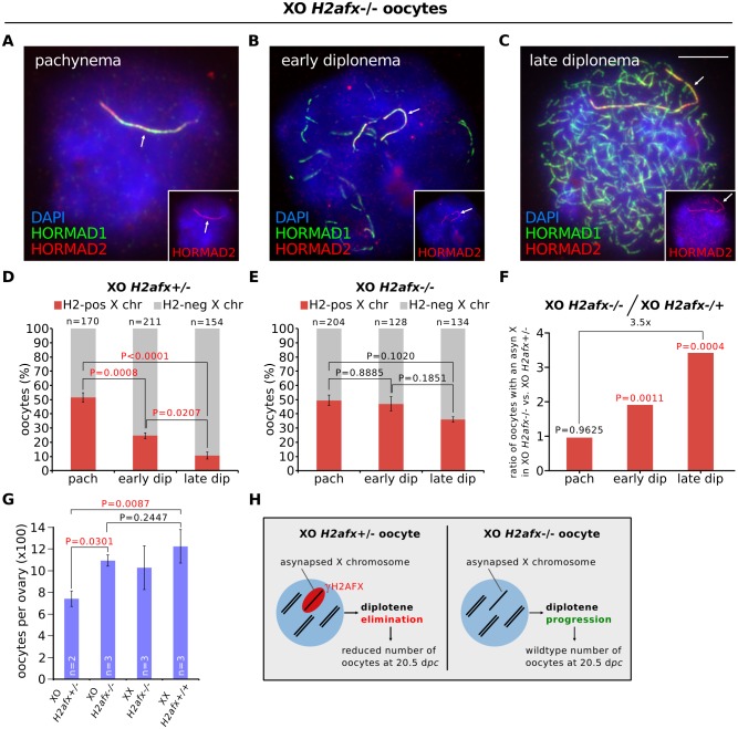
Fig 3. H2afx deletion reverses perinatal oocyte losses in XO females.
(A) Pachytene XO H2afx-/- oocyte with an asynapsed X chromosome (arrow; marked by HORMAD1, green, and HORMAD2, red and inset). (B) Early diplotene XO H2afx-/- oocyte, showing intermediate levels of desynapsis (HORMAD1-only positive axes, green) and an asynapsed X chromosome (arrow; marked with both HORMAD1 and HORMAD2, inset). (C) Late diplotene XO H2afx-/- oocyte, showing extensive desynapsis and an asynapsed X chromosome (arrow). Scale bar represents 10μm. (D) Mean percentage (± s.e.m.) of XO H2afx+/- oocytes with a HORMAD1/2 double-positive X chromosome between pachynema and late diplonema at 19.5 dpc. Statistics were performed as in Fig 1. (E) Mean percentage (± s.e.m.) of XO H2afx-/- oocytes with a HORMAD1/2 double-positive X chromosome between pachynema and late diplonema at 19.5 dpc. No statistically significant difference was found between means at pachynema and late diplonema (Tukey’s test). (F) Enrichment of oocytes with an asynapsed X chromosome in XO H2afx-/- compared to XO H2afx+/- between pachynema and late diplonema. Enrichment was defined as the ratio of the mean percentage of oocytes with an asynapsed X in XO H2afx-/- versus XO H2afx+/- control, using data from part (D) and (E). (G) Mean number (± s.e.m.) of oocytes in XO and XX, ovaries with varying doses of H2afx, XO H2afx-/-,XX H2afx+/- and XX H2afx+/+ at 20.5 dpc. n is the number of non-littermate ovaries analyzed. Tukey multiple comparison tests. (H) Summary showing the importance of H2AFX in XO oocyte elimination.

