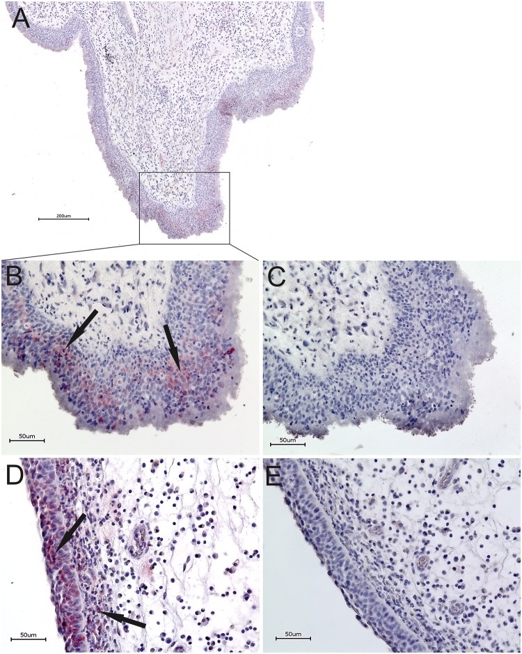
Fig 2. HPV Immunohistochemistry.
IHC Overview of a representative HPV-positive antrochoanal polyp with HPV16 staining (A). HPV16 E6 protein staining of a representative HPV-positive antrochoanal polyp, relevant epithelial region shown (B) with corresponding negative control (C). HPV16 E6 protein staining of a representative HPV-positive nasal polyp, relevant epithelial region shown and subepithelial stained cells (D) with corresponding negative control (E). Discontinuous epithelial cell clusters stained positive in HPV-positive antrochoanal- and nasal polyps.

