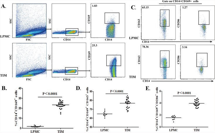
Fig 2. Characterization of CD14+CD169+ macrophages in the colorectal tissues.
A total of 30 fresh surgical CRC tumor tissues were digested for characterization of tumor infiltrating macrophages (TIMs). In addition, 12 lamina propria tissues from non-tumor patients were digested for characterization of macrophages in total lamina propria mononuclear cells (LPMCs). Subsequently, the cells were stained with anti-CD14, CD169 and CD163 or CD206. The frequency of CD14+CD169+, CD14+CD169+CD163+ and CD14+CD169+CD206+ macrophages in LPMCs and TIMs was determined by flow cytometry. Data are representative charts or expressed as the values of individual subjects. The horizontal lines indicate the median for individual groups. (A) Flow cytometry analysis of CD14+CD169+ in LPMCs and TIMs. (B) The percentages of CD14+CD169+ macrophages in LPMCs and TIMs. (C) Flow cytometry analysis of CD14+CD169+CD163+ and CD14+CD169+CD206+ macrophages in total CD14+CD169+ LPMCs and TIMs macrophages. (D) The frequency of CD14+CD169+CD163+ macrophages in LPMCs and TIMs. (E) The frequency of CD14+CD169+CD206+ macrophages in LPMCs and TIMs.

