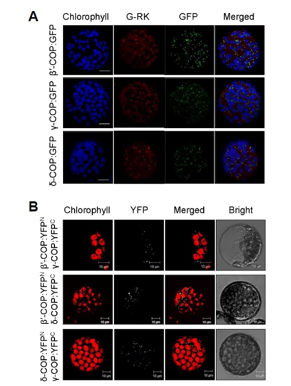
Fig. 1.

Subcellular localization and protein interactions of COPI subunits. Each experiment was performed at least three times, and representative images are shown. (A) GFP-fusion proteins of N. benthamiana β′-, γ-, and δ-COP were expressed in N. benthamiana leaves through agroinfiltration, and GFP signal in leaf protoplasts was examined by confocal microscopy. To mark the Golgi complex, G-RK (red fluorescence) was co-expressed with the GFP fusion proteins. Chlorophyll autofluorescence is pseudo-colored blue. Scale bars = 10 μm. (B) BiFC was performed to detect in vivo interactions between N. benthamiana COPI subunits. β′-COP:YFPN and γ-COP:YFPC, β′-COP:YFPN and δ-COP:YFPC, and δ-COP:YFPN and γ-COP:YFPC were expressed together via agroinfiltration, and the YFP signal in leaf protoplasts was observed by confocal microscopy.
