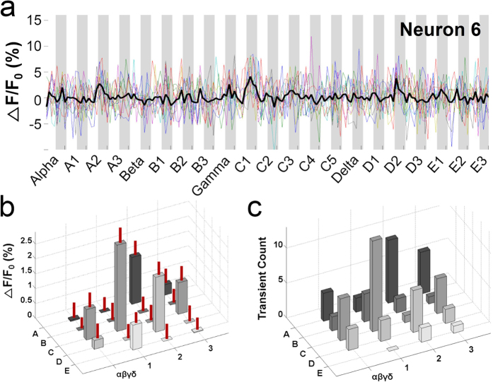
Figure 4. A neuron with broad whisker tuning.
(a) Averaged Ca2+ signals of Neuron 6 in Fig. 2b in response to stimulation of 21 whiskers. (b) Topographical 3D plot of Ca2+ signals against stimulated whiskers. The S.E.M. is indicated as a vertical red line on each bar. This neuron was tuned not only to the C1 whisker but also to other whiskers, shown as multiple peaks in Ca2+ responses (one-way ANOVA, F(18,741) = 4.342, p < 0.001; Holm-Sidak method for multiple comparisons between the C1 response and the other responses; α: p = 0.003; A1: p < 0.001; A2: p = 0.996; A3: p = 0.021; β: p < 0.001; B1: p < 0.001; B2: p = 0.001; B3: p < 0.001; γ: p = 0.501; C2: p < 0.001; C3: p = 0.554; δ: p = 0.015; D1: p = 0.002; D2: p = 1; D3: p < 0.001; E1: p = 0.244; E2: p < 0.001; and E3: p = 0.003). (c) Topographical 3D plot of transient counts against stimulated whiskers.

