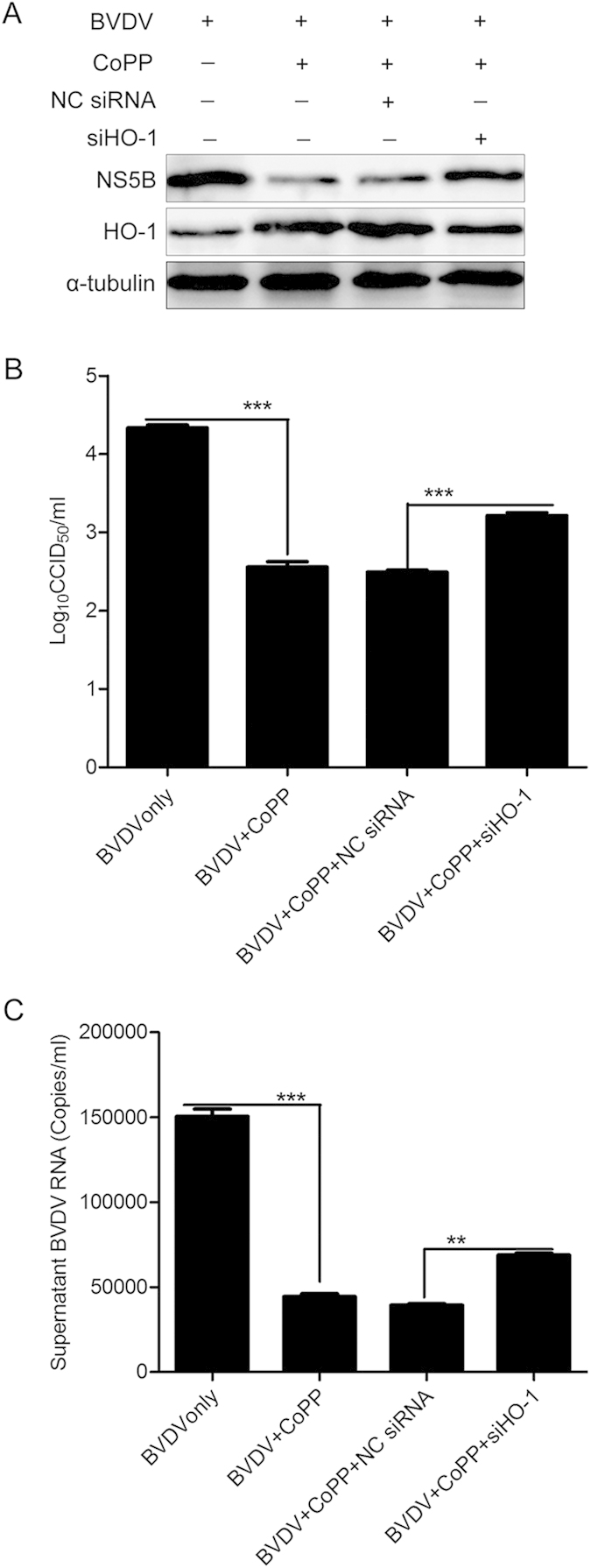
Figure 7. CoPP-induced attenuation of BVDV is HO-1 dependent.

siRNAs specific to HO-1 were transfected into MDBK cells, which were then treated with CoPP for 12 h. And then, these cells were infected with BVDV at a MOI of 0.1 for 36 h. Subsequently, the intracellular HO-1 and BVDV NS5B protein expression levels were analyzed by Western blot (A), and α-tubulin was used as the control. Virus titers (B) and BVDV RNA (C) in the supernatants were analyzed by CCID50 and qRT-PCR. Data are expressed as mean ± s.d. of three independent experiments, **P < 0.01, ***P < 0.001.
