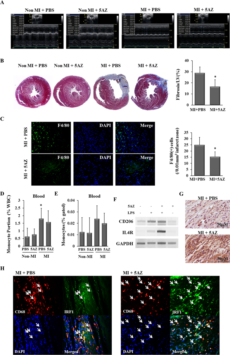
Figure 4. 5AZ improves LV function and regulated the phenotype of infiltrated macrophages.
(A) Representative echocardiograms showed more preserved cardiac function in the MI + 5AZ group than in the MI + PBS group at 2 weeks after MI. (B) Cardiac fibrosis was examined by Masson’s trichrome staining and quantified. *Fibrotic area was significantly smaller in the MI + 5AZ group than in the MI+PBS group, p < 0.05. (C) The representative images of infiltrated F4/80(+) macrophages in the infarcted myocardium were shown and the number of macrophages was expressed in a graph (n = 4 per group). (D) Circulating monocytes were counted by complete blood count and the percent of WBC was expressed. *The portion of circulating monocyte was significant higher than other three groups, p < 0.05 (E) CD45(+)CD14(+) monocytes from whole blood were quantified by flow cytometry and the percent of monocytes of CD45(+) cells was expressed. (F) The mRNA levels of CD206 and IL4R, M2 macrophage markers, were potentiated by 5AZ in LPS-stimulated RAW264.7 cells. (G) Immunohistochemical staining for the anti-inflammatory M2 macrophage marker CD206 in mouse infracted myocardium. CD206(+) cells were more frequently observed in the MI+5AZ group than in MI + PBS group. (H) IRF1-expressing macrophages in the infarcted myocardium. Double immunohistochemical staining for identification of IFR-1 expression in infiltrated macrophages in rat infarcted myocardium. CD68(+) IRF1(+) cells were abundant in the MI+ 5AZ group compared with the MI+PBS group. White arrows indicate the CD68(+) IRF1(+) cells.

