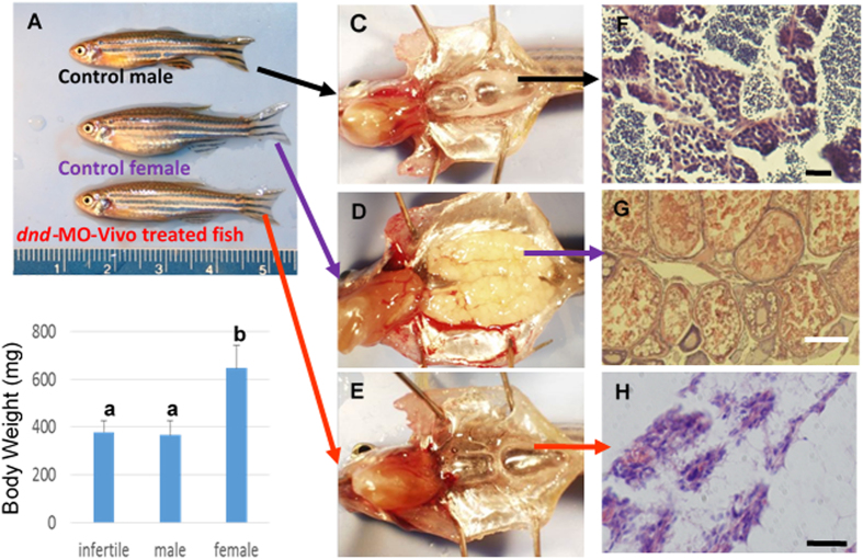
Figure 3. Zebrafish dnd-MO-Vivo induced sterility in zebrafish.
Embryos initially treated with 60 or 40 μM of dnd-MO-Vivo developed into male-like adults. (A) No difference in appearance or overall size was observed between treated adult fish and control males. (B) No significant difference in body-weight (Mean ± SD) of 3-month-old fish (N = 12 by random sampling) was noted among dnd-MO-Vivo treated fish and control males (Data that share the same letter are not significantly different from each other). Examination of gonadal tissue showing (C) a fully-developed testis of a control male fish, (D) a fully-developed ovary of a control female fish, (E) the gonads of dnd-MO-Vivo treated fish that developed into a thin filament-like tissue. Photomicrographs (F–H) show (F) advanced spermatogenesis in the testis of a control male fish, (G) a well-developed ovary of a control female fish with oocytes at advanced stages of gametogenesis, (H) the gonad of dnd-MO-Vivo treated fish appears to be under-developed and surrounded with a large amount of adipocytes, without advanced gonadal structure or germ cells. Scale bar: white = 200 μm, black = 20 μm.

