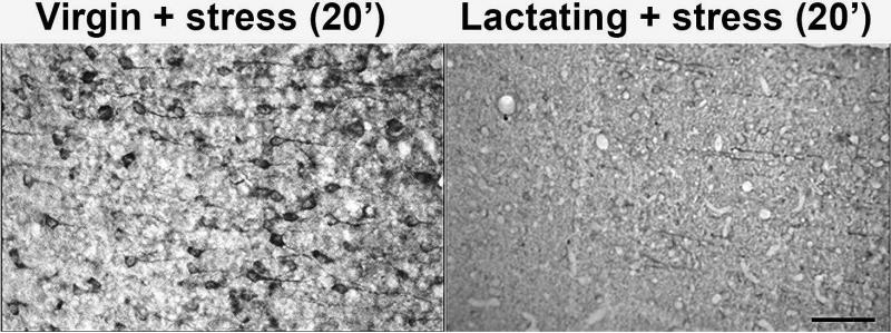
Figure 5. Localization of tau-P in the CA3 region of stressed virgin or lactating rat hippocampus.
30um thick sections of the hippocampus were stained with PHF-1 to visualize the localization of tau-P in stressed virgin and lactating rats twenty minutes following stress (N=5, each). This 20x magnification of CA3 pyramidal neurons demonstrates a lack of increase in somatic staining in stressed lactating animals (right) compared to stressed virgin animals (left). Unstressed controls (not shown) had no appreciable tau-P signal. Stressed lactating animals did not significantly differ in tau-P levels compared to unstressed controls (p>0.05). Bar = 100μm.

