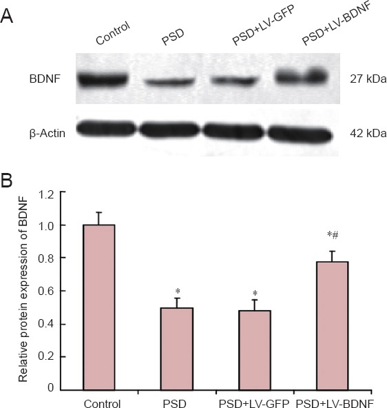
Figure 4.

BDNF protein expression in rat hippocampus at 7 days post injection of lentivirus by western blot assay.
(A) Molecular weight of BDNF is 27 kDa, and β-actin protein is 42 kDa. Bands for the four groups are shown (control, PSD, PSD + LV-GFP and PSD + LV-BDNF). (B) The gray value ratio of the BDNF band to the β-actin band was calculated, and the relative expression was compared with the control group. *P < 0.05, vs. control group; #P < 0.05, vs. PSD + LV-GFP group. The data are presented as the mean ± SEM with five rats in each group. Statistical analysis was performed using one-way analysis of variance and Tukey's post hoc test. PSD: Post-stroke depression; LV: lentivirus; GFP: green fluorescent protein; BDNF: brain-derived neurotrophic factor.
