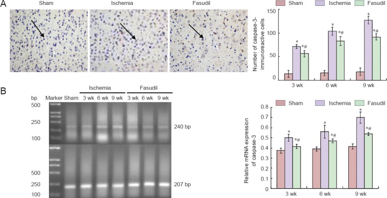
Figure 6.

Immunoreactivity and mRNA expression of caspase-3 in the frontal cortex of rats following chronic cerebral ischemia and the effects of fasudil treatment.
(A) Left: Representative photomicrographs of caspase-3-immunoreactive cells (arrows) in rats in the sham and ischemia groups at 3, 6, 9 weeks (wk) after surgery. Right: Quantification of caspase-3 immunoreactivity. (B) mRNA expression of caspase-3. The mRNA results are expressed as the ratio of the target gene band intensity to that for β-actin. There were 10 rats at each time point per group. Data are expressed as the mean ± SD and analyzed using one-way analysis of variance and the Student's t-test. *P < 0.05, vs. sham operated (sham) group; #P < 0.05, vs. ischemia group.
