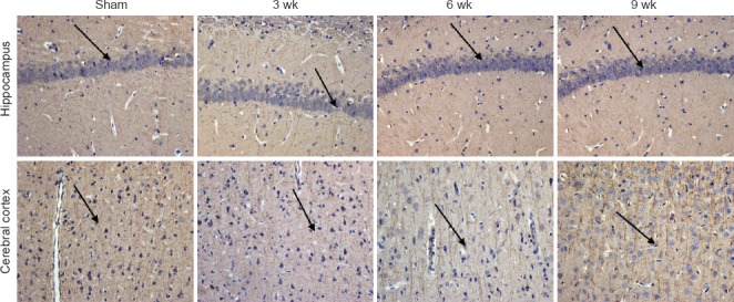
Figure 7.

Representative photomicrographs of microtubule-associated protein 2 (MAP2)-immunoreactive cells in the hippocampus and frontal cortex of rats following chronic cerebral ischemia (200×).
MAP2-immunoactive cells in rats in the sham and ischemia groups at 3, 6, 9 weeks after surgery. Arrows indicate MAP2-immunoreactive cells.
