
Figure 1.
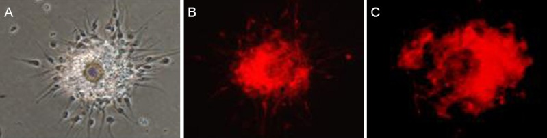
Culture, identification, and labeling of neural stem cells (NSCs).
(A) Morphology of primary NSCs by inverted phase contrast microscopy (× 40); (B) NSCs positive for Nestin (immunofluorescence staining, × 40, red); (C) CM-Dil-positive NSCs (× 100; red).