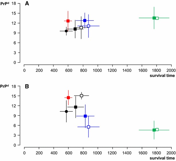
Figure 5.

Accumulation of PrP d in CNS and LRS tissues in relation to survival times. Magnitude of PrPd accumulation in the brain (A; Y axis as mean ± SD of individual sheep scores for total PrPd (=sum of scores in seven standard brain areas)) and in LRS tissues (B; Y axis as mean ± SD of individual sheep scores for total PrPd (=sum of scores in six different lymphoid tissues)) of sheep clinically affected with BSE after oral (solid symbols) or natural (open symbols) transmission in relation to their survival times (X axis in days, mean ± SD). Black, ARQ Suffolk sheep (circle, 1 g dose; square, 5 g dose); blue, ARQ Romney sheep; green, VRQ Cheviot sheep; red, AHQ Cheviot sheep. For number of animals in each group refer to Table 1. Note that (1) there is significant individual variation in PrPd scores in both brain and LRS tissues, (2) brain PrPd scores are relatively similar between groups regardless of differences in survival time and (3) LRS scores are low for VRQ Cheviot sheep despite their long survival time and high brain PrPd.
