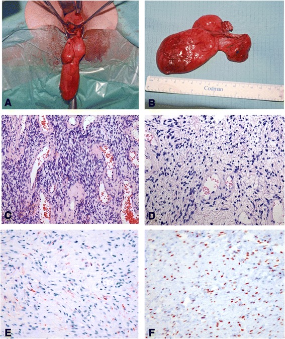
Fig. 3.

a and b Intraoperative appearance of the tumor. The tumor was covered entirely with a frail membrane of pinkish gray appearance. c Cellular mesenchymal lesion with alternating cellularity intermingled with small blood vessels [hematoxylin and eosin (H&E) stain, original magnification ×109]. d Higher-magnification image representing thin-walled blood vessels surrounded by ovoid to spindle-shaped cells with some epithelioid appearance and abundant eosinophilic cytoplasm (H&E stain, original magnification ×241). e Immunohistochemical staining for desmin with weak positivity. f Strong nuclear expression of estrogen receptor.
