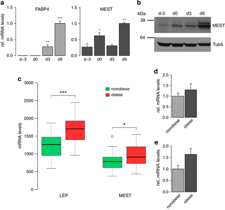
Figure 1.
Endogenous expression of MEST in human adipocyte differentiation and adipose tissue. (a and b) hMADS cells were grown to confluence (d-2) and stimulated to undergo adipocyte differentiation 2 days later (designated d0). (a) RNA was prepared at indicated time points and subjected to quantitative real-time reverse transcription PCR (RT-qPCR) for fatty acid-binding protein 4 (FABP4) and MEST. Expression levels were normalised to TBP and are presented relative to d9 (n=3). *P<0.05, **P<0.01, ***P<0.001 versus undifferentiated cells at d-3. (b) Protein lysates were prepared at indicated time points and analysed by western blotting for MEST and β-tubulin (Tubβ). (c) Abdominal scWAT biopsies from 26 non-obese and 30 obese female donors were analysed by microarray (Gene Expression Omnibus experiment GSE25401). Expression levels of leptin (LEP) and MEST are shown as boxplots. *P<0.05, ***P<0.001. (d and e) Expression of MEST was analysed by RT-qPCR in (d) adipocyte progenitor cells (CD45−/CD31−/CD34+) from scWAT biopsies of 10 non-obese and 8 obese subjects, and (e) mature adipocytes from scWAT biopsies of 4 non-obese and 6 obese subjects. MEST expression levels were normalised to 18S rRNA.

