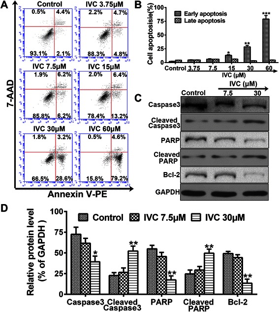
Fig. 3.

IVC induces apoptotic death. a MHCC97H cells were incubated with IVC at indicated doses for 24 h, and Annexin V-PE and 7-AAD stained cells were sorted by flow cytometry. b Distribution of cell apoptosis was analyzed. c Protein expressions of Caspase3, Cleaved Caspase3, PARP and Bcl-2 were detected by western blot. d The relative protein level in each condition was quantitated using Image J. Experiments were repeated three times, and similar results were obtained. Data are expressed as mean ± S.E. *P < 0.05, **P < 0.01 and ***P < 0.001 vs. the control
