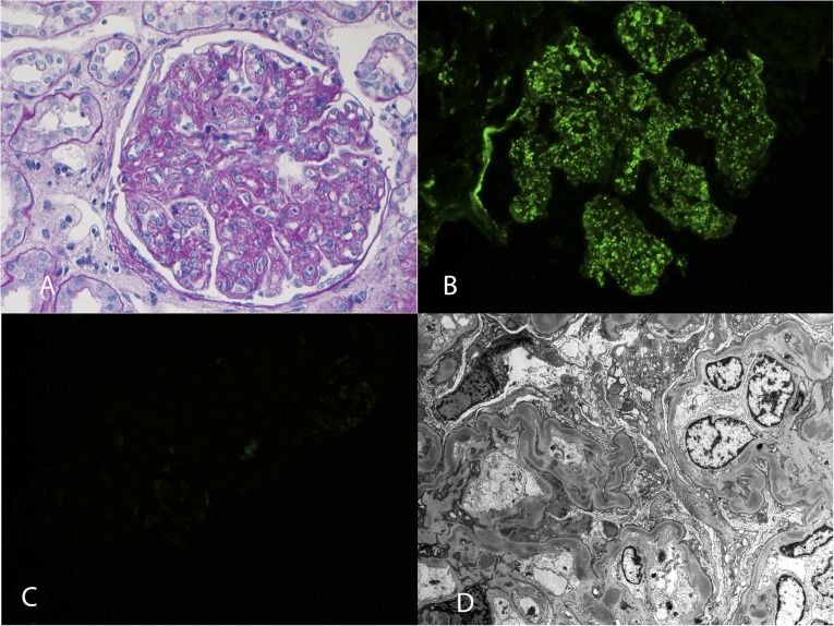
Figure 3.
DDD. (A) Periodic acid–Schiff stain showing MPGN with mesangial expansion, increased mesangial cellularity, thickened capillary walls, and double contour formation (40×). Immunofluorescence showing (B) bright staining for C3 in the mesangium and along the capillary walls (40×) and (C) completely negative staining for C4d. (D) Electron microscopy showing dense deposits along the glomerular basement membranes and in the mesangium (2900×).

