Figure 6.
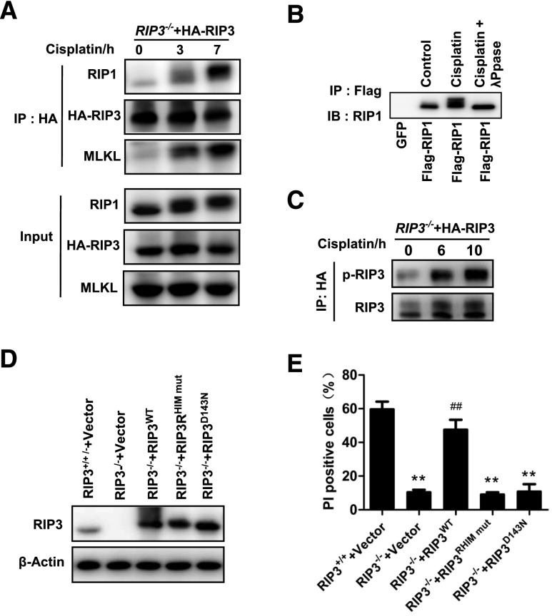
The formation of necrosomes containing RIP1, RIP3, and MLKL is required for cisplatin-induced cell necroptosis in PTCs. (A) PTCs from RIP3−/− mice were infected with HA-RIP3–expressing lentivirus before stimulation with 100 µM cisplatin for the indicated time (n=3). Cell lysates were immunoprecipitated with anti-HA antibody (IP: HA) and analyzed by immunoblotting with anti-RIP1, anti-RIP3, or anti-MLKL antibodies. Input, 5% of extract before immunoprecipitation (control). (B) PTCs were infected with a lentivirus encoding green fluorescent protein or Flag-RIP1 before stimulation with 100 µM cisplatin for 4 hours (n=3). Immunoprecipitations were performed using M2 beads. The immunoprecipitates were treated with or without λ-phosphatase, and then analyzed by immunoblotting with anti-RIP1 antibody. (C) PTCs from RIP3−/− mice were infected with HA-RIP3–expressing lentivirus and then stimulated with 100 µM cisplatin for the indicated time period (n=3). Cell lysates were immunoprecipitated with anti-HA antibody (IP: HA) and analyzed by immunoblotting with anti-RIP3 or anti–P-RIP3 antibodies. (D and E) Cells from RIP3−/− mice were infected with a lentivirus encoding nothing (vector) or Flag-RIP3wt, Flag-RIP3RHIM mut, or Flag-RIP3D143N for 36 hours. Cells from RIP3+/+ mice were infected with empty vector. RIP3 expression levels are shown in D by Western blot. All cells were stimulated with 100 µM cisplatin for 12 hours before quantification of PI-positive cells and shown in E (n=4). **P<0.01 versus RIP3+/++vector group; ##P<0.01 versus RIP3−/−+vector group.

