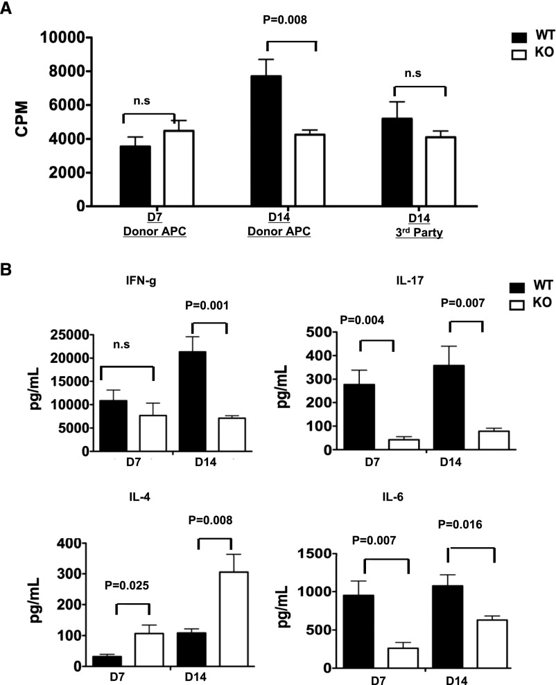
Figure 4.
T cells from MyD88−/− recipient mice display decreased proliferative capacity and effector function at 14 days, but not 7 days, after transplantation. (A) MLRs were set up using T cells isolated from the spleens of WT or MyD88−/− kidney allograft recipients 7 or 14 days after transplantation. Responding T cells were stimulated with donor B6 APCs or third-party SJL APCs as indicated. Proliferation was assessed by [3H] thymidine uptake during the last 18 hours of a 5-day MLR. (B) Culture supernatants from the MLR stimulated with donor APCs as in part A were collected immediately before thymidine pulse and analyzed for the presence of IFN-γ, IL-4, IL-6, and IL-17 by Luminex bead assay. Results represent two independent experiments with two to three mice per group. KO, knockout.

