Figure 2.
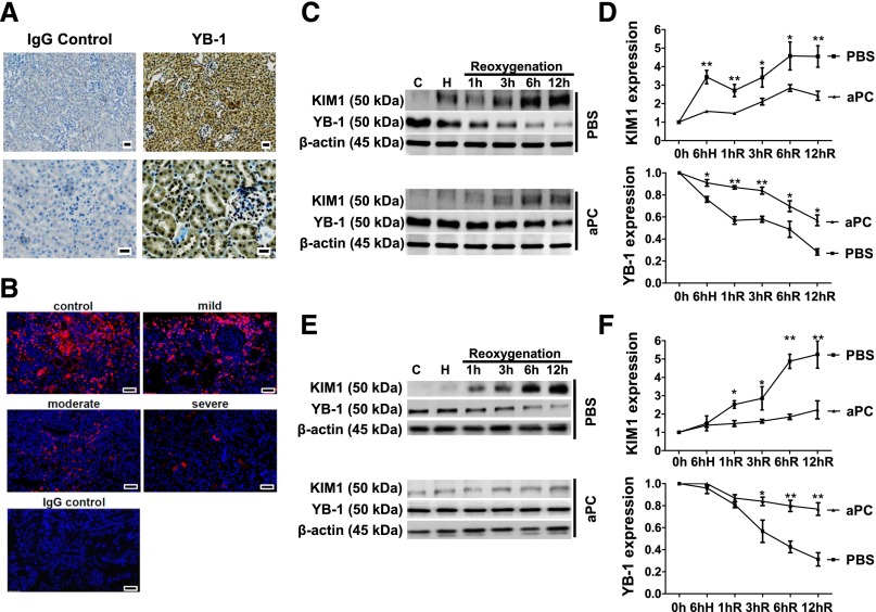
aPC maintains YB-1 expression in tubular cells after HR. (A) YB-1 is strongly expressed in renal tubular cells. Exemplary immunohistochemical YB-1 staining of renal paraffin-embedded tissue sections from healthy WT mice (right), IgG control (left), and YB-1 antigen detected by HRP-DAB reaction (brown) and hematoxylin counterstain (blue); overview (top) and tissue section at higher magnification (bottom); scale bar: 20 µm. (B) Expression of YB-1 (red) in human renal biopsies is reduced after acute renal injury. Exemplary immunofluorescent staining of renal paraffin-embedded tissue biopsies from patients with AKI graded as mild, moderate, or severe and control tissue sections; scale bar: 20 µm. Time-dependent expression of KIM1 and YB-1 in mouse tubular cells (BUMPT) [(C) and (D)] and in primary renal proximal tubular epithelial cells (rTEC) [(E) and (F)] in vitro at baseline, after 6 hours of hypoxia (H) (1% O2 for 6 hours), and at various time points (1–12 hours) after reoxygenation (21% O2). Pretreatment with aPC (20 nM) diminishes the increase of KIM1 and the loss of YB-1 expression compared with PBS-treated control cells; representative immunoblots of whole-cell lysates [(C) and (E)] and line graph [(D) and (F)] summarizing results. Mean±SD value of at least three independent experiments [(D) and (F)]; *P<0.05; **P<0.01 (ANOVA).

