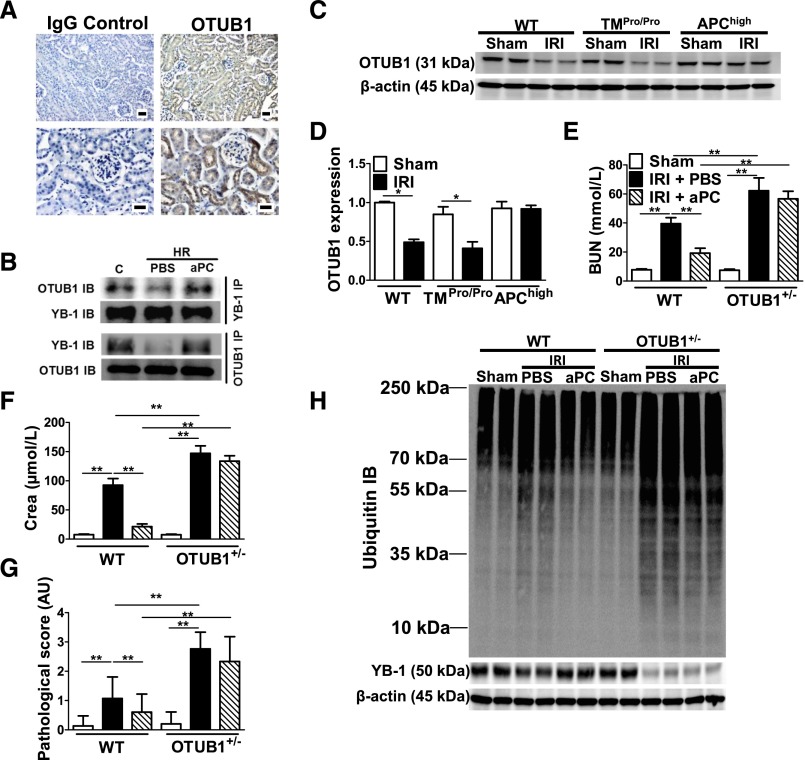
Figure 5.
aPC-mediated renal protection and sustained YB-1 expression after IRI depends on OTUB1. (A) OTUB1 is predominately expressed in tubular cells; immunohistochemical detection of OTUB1 in paraffin-embedded tissue sections of WT mouse kidney (right) and IgG control (left); OTUB1 antigen detected by HRP-DAB reaction (brown) and hematoxylin counterstain (blue); overview (top) and tissue section at higher magnification (bottom); scale bar: 20 µm. (B) The interaction of YB-1 and OTUB1 is impaired after HR injury in tubular cells. Detection of OTUB1 by immunoblotting after immunoprecipitation of YB-1 (top; YB-1 IB: input control) or of YB-1 after immunoprecipitation of OTUB1 (bottom; OTUB1 IB: input control) from whole-cell lysates of control (C) mouse tubular cells or tubular cells after in vitro HR without (PBS) or with aPC treatment (aPC) (20 nM); representative images of immunoblots. Expression of OTUB1 is maintained in APChigh mice after IRI. OTUB1 protein levels in kidney lysates were determined by immunoblotting and normalized to β-actin; representative immunoblots (C) and bar graph summarizing results (D). BUN (E), creatinine (Crea) (F), and tissue injury (pathologic score) (G) in control (Sham, open bars) and experimental mice without (IRI) (black bars) or with aPC pretreatment (IRI+aPC) (striped bars; aPC: 0.5 mg/kg i.p.). (H) Immunoblot of ubiquitinated proteins (top) and YB-1 (middle) and β-actin as loading control (bottom) in WT and heterozygous OTUB1 (OTUB1+/−) mice. Control mice (Sham) or mice with IRI without (PBS) or with (aPC) (0.5 mg/kg i.p.) treatment. Mean±SD value of at least six mice per group [(D)–(G)]; *P<0.05; **P<0.01 (ANOVA).

