Figure 2.
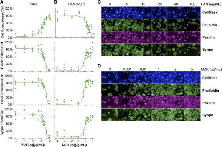
The novel assay quantitatively measures phenotypic changes in podocytes. (A) PAN induces dose–dependent podocyte damage. Podocytes in 96-well optical plates were treated with an increasing dose of PAN at 37°C for 48 hours, and the cellular damage was assessed on staining podocytes with CellMask Blue (to measure cell morphology), phalloidin (to quantify F-actin fibers), anti-paxillin antibody (to quantify focal adhesions), and anti-synaptopodin (anti-synpo) antibody (to quantify synaptopodin levels) and analyzing them using the newly developed HCS assay. Dose-response curves showing the effects of increasing concentrations of PAN on four different cellular parameters (cell morphology [as defined by cell roundness], the number of actin fibers per cell, the number of focal adhesions per cell, and the number of synpo fibers per cell) as a way to measure PAN–induced podocyte injury. The x axis represents PAN concentration, and the y axis shows quantification of each of four parameters at a defined dose of PAN. Data shown are means±SEMs per cell from a single-assay well (n=500–1000 cells) performed in three replicate wells. (B) MZR dose dependently protects podocyte from PAN injury. Podocytes in 96-well optical plates were cotreated with PAN (30 μg/ml) and an increasing concentration of MZR at 37°C for 48 hours, and the cellular damage was assessed using an HCS system. Dose-response curves showing the protective effects of increasing concentration of MZR on various cellular parameters (as with PAN treatment) are presented. Data shown are means±SEMs per cell from a single-assay well (n=500–1000 cells) performed in three replicate wells. (C) Representative fluorescence images of cells treated with increasing doses of PAN (as shown), stained with CellMask Blue, phalloidin, antipaxillin antibody, or antisynpo antibody, and quantified as shown in A. Scale bar, 50 μm. (D) Representative fluorescence images of cells cotreated with PAN (30 μg/ml) and an increasing dose of MZR (as shown), stained with CellMask Blue, phalloidin, anti-paxillin antibody, or anti-synpo antibody, and quantified as shown in B. Scale bar, 50 μm.

