Figure 6.
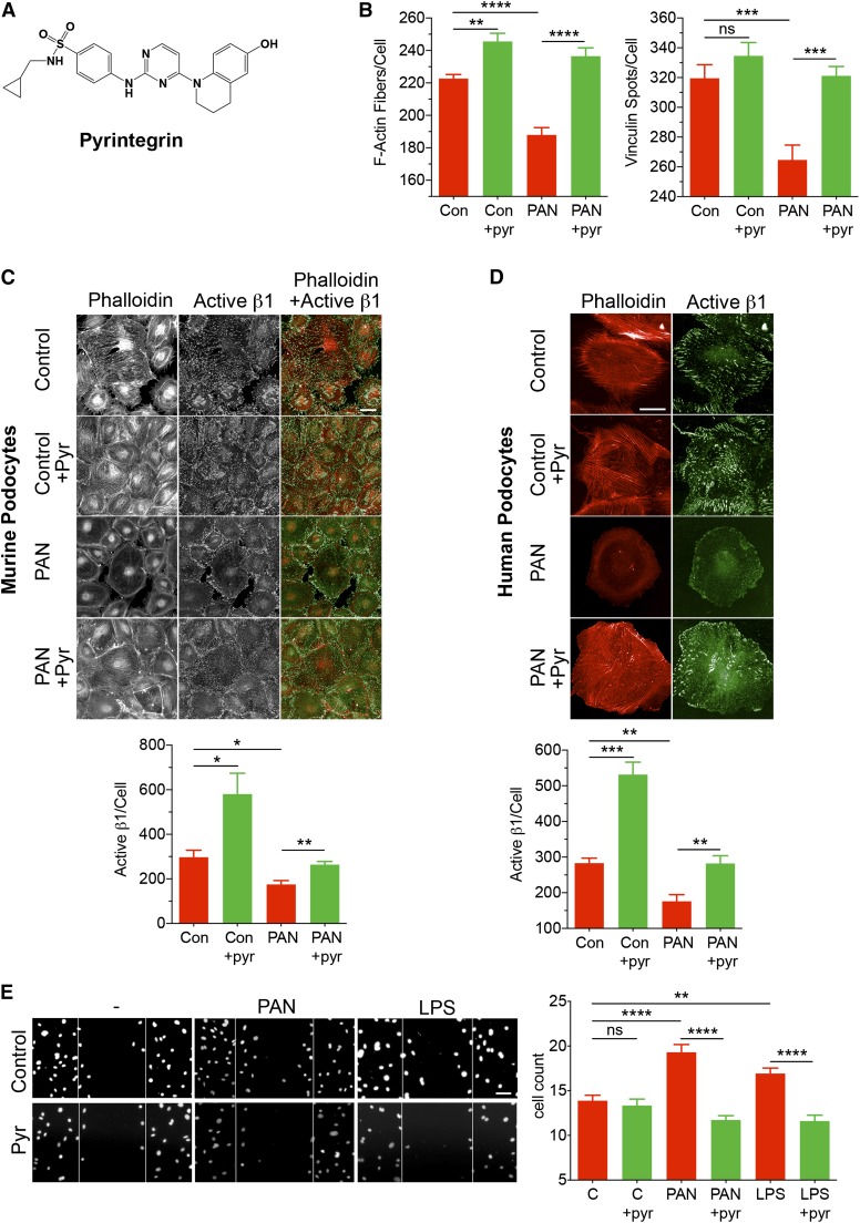
An β1-integrin agonist pyrintegrin (pyr) protects podocytes from damage. (A) The published chemical structure of pyr.34 (B–D) Pyr protects podocytes from PAN-induced loss of F-actin fibers and focal adhesions and enhances active β1-integrin levels. Podocytes in 96-well optical plates were cultured at 37°C for 48 hours in the absence (control [Con]) or presence of PAN (30 μg/ml) and cotreated with vehicle (DMSO; 1%) or pyr (1 μM). The cellular damage was assessed after staining the cells with phalloidin, anti-vinculin, and anti-active β1-integrin antibodies and quantified using the HCS system. (B) Graphs showing the effect of pyr on the number of F-actin fibers per cell and the number of vinculin spots per cell under each treatment condition. Data shown are means±SEMs per cell from three replicate wells (n=500–1000 cells per well). **P<0.01; ***P<0.001; ****P<0.001. (C) Representative fluorescence images of murine podocytes after various treatments and after staining with phalloidin and antiactive β1 antibody. Two–color costained images show cells stained with phalloidin (red) and anti-active β1 antibody 9EG7 (green). Images were acquired using the Opera HCS System. The graph (lower panel) shows quantification of the active β1-integrin spot intensity per cell under each treatment condition. Data shown are means±SEMs per cell from four to five replicate wells (n=500–1000 cells per well). Scale bar, 50 μm. *P<0.01; **P<0.001. (D) Representative fluorescence images of human podocytes after various treatments as shown. Images show cells stained with phalloidin (red) and anti-active β1-integrin antibody 12G10 (green). Images were acquired using the Opera HCS System. The graph (lower panel) shows quantification of the active β1-integrin spots per cell under each treatment condition. Data shown are medians±SEMs per cell from four to five replicate wells (n=500–1000 cells per well). Scale bar, 50 μm. **P<0.01; ***P<0.001. (E) Pyr reduces injury-mediated increase in podocyte migration in a scratch wound–healing assay. Representative images (left panel) and a bar graph (right panel) showing confocal microscopy–based quantitation of the number of migrating podocytes in a scratch wound–healing assay. Wounds were created in podocyte monolayers using sterile pipette tips, and the cells were incubated in the absence (−) or presence of PAN or LPS at 37°C for 48 hours. One set of wounds was cotreated with pyr. Subsequently, cells were fixed and stained with 4′,6-diamidino-2-phenylindole (DAPI), and the cells migrating inside the edge of the wounds were quantified. Lines represent the wound margins at the beginning of the experiment (0 hours). The data presented in the graph are plotted as means±SEMs (n≥15). C, control. Scale bar, 50 μm. **P<0.01; ****P<0.001.

