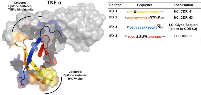
Fig. 3.

Structure of the IFX Fab fragment interaction with TNF-α. The identified IFX epitopes are located in or in close proximity to the IFX CDR involved in TNF-α binding. The mapped epitopes of the IFX variable segment of the heavy chain are indicated in yellow and orange (IFX 1 and 2), the light chain epitopes are shown in blue and red (IFX 3 and 4). The amino acids with direct contact to TNF-α in the CDR of IFX are indicated by black letters in the epitope sequences. Boxed N41 in epitope IFX 3 is part of a glycosylation sequon
