Fig. 4.

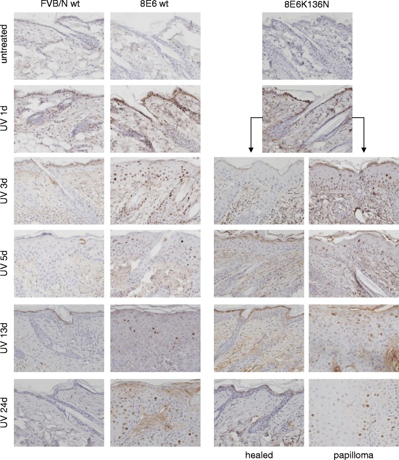
Presence of DNA damage in UV treated skin of K14-HPV8-E6 mice. Paraffin embedded skin sections of UV treated skin were stained for γH2AX (magnification: 400×). Representative images of n = 4 mouse skin biopsies per time-point are shown

Presence of DNA damage in UV treated skin of K14-HPV8-E6 mice. Paraffin embedded skin sections of UV treated skin were stained for γH2AX (magnification: 400×). Representative images of n = 4 mouse skin biopsies per time-point are shown