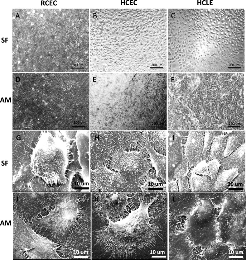
Figure 3.
Phase-contrast images of corneal epithelial cells cultured on (A–C) SF and (D–F) AM showing comparable cell morphology; SEM images of corneal epithelial cells on (G–I) SF and (J–L) AM by using three different cell sources: (A, D, G, J) RCEC, (B, E, H, K) HCEC, and (C, F, I, L) HCLE demonstrating a high level of cell anchor point to the film surface, abundance of microvilli on the cell surface, and prevalent contacts between adjacent cells.

