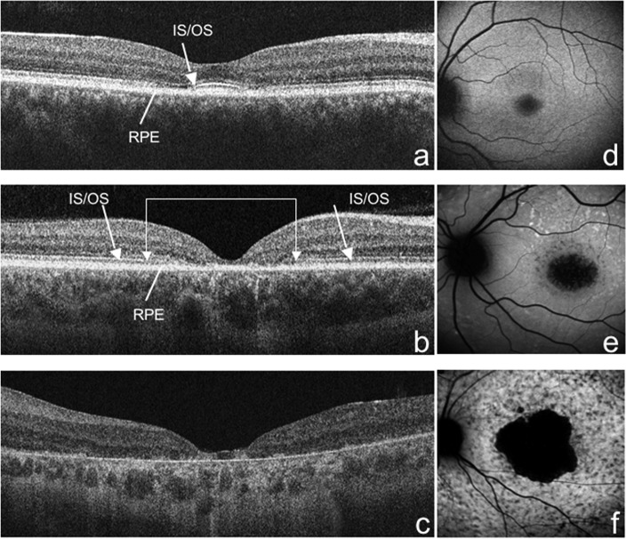
Figure 1.
SD-OCT. (a) IS/OS junction disorganization in the fovea (arrow); (b) IS/OS junction loss closer to the fovea (double arrow) compared with the extrafoveal area (arrows); (c) extensive loss of IS/OS junction and RPE atrophy. (d) FAF; presence of a ring of increased autofluorescence surrounding an area of decreased autofluorescence; (e) absence of foveal autofluorescence (<1 disc diameter); and (f) absence of macular autofluorescence (≥2 disc diameter).

